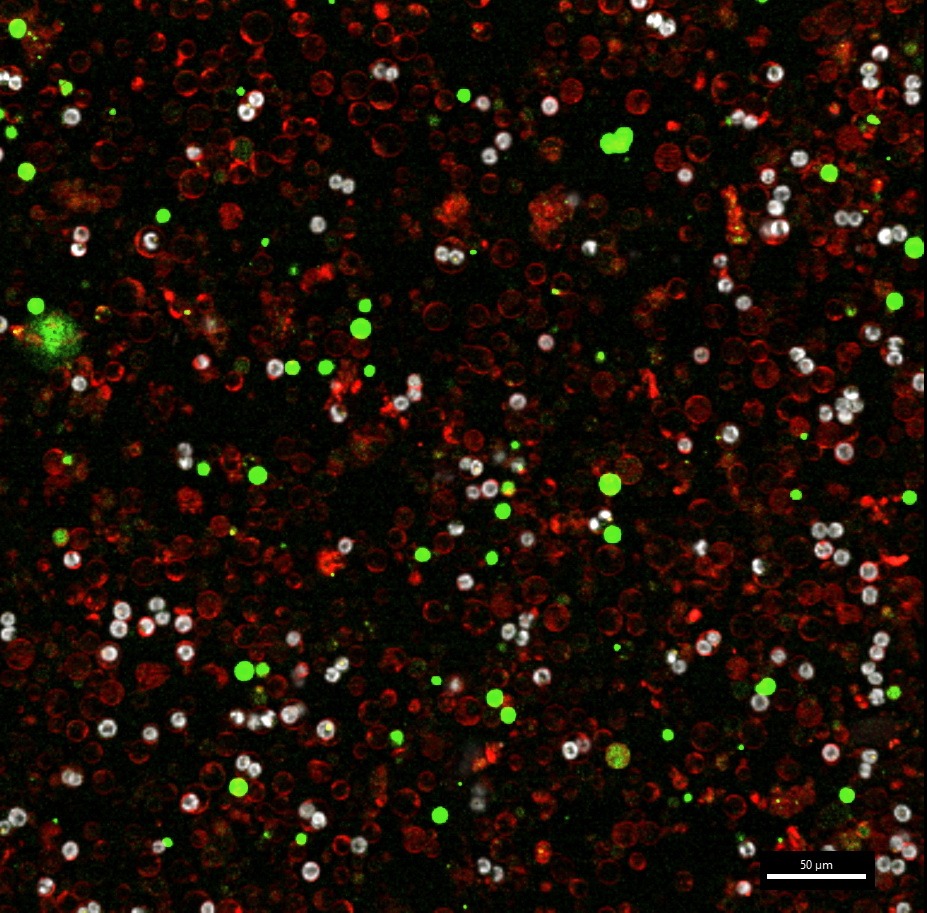
Confocal image of cells dissociated from coral tissue. Coral cells expressing native green fluorescent protein (GFP, green); algae symbionts (chlorophyll autofluorescence, white) reside within coral gastrodermal cells (FM4-64, red).
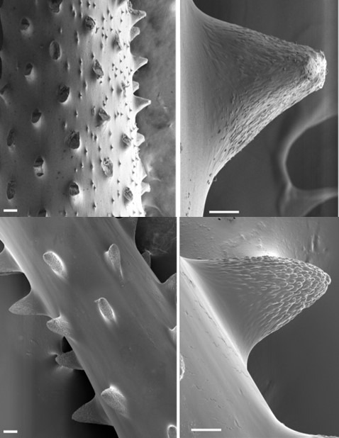
Black Corals (Antipatharia)
Dr. Miri Morgulis/ Prof. Tali Mass Lab
Scanning electron microscopy (SEM) image of skeletal morphology of the two northern Red Sea black coral species. Horizontal cross section of the skeleton showing primary branchlets and spines (left panel, scale bar: 100 μm). Spines magnification (right panel, scale bar: 20 μm).
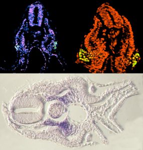
Catshark embryo
Pascal Schmidt/ Dr. Ram Reshef Lab
Wide-field fluorescence (top) and Bright-field (bottom) images of cross sections of catshark embryos at the developmental stage of early kidney formation. Upper left: PCNA, cell proliferation marker (red) and Pax2, a kidney marker (green), DAPI (nuclei, blue). Upper right: PCNA (red) overlaps with Pax2 (green/yellow) in the budding tissue of the kidney. Bottom: expression of Pax1, the sclerotome marker (bone formation), in the catshark somite.
Sea Urchin Embryo
Dr. Tsvia Gildor/ Prof. Smadar Ben-Tabou de-Leon Lab
Confocal image of sea urchin embryo, 24 hours post fertilization. F-actin filaments (red). Skeletogenic cells (green) showing the enrichment of actin filaments around the embryo’s skeleton.
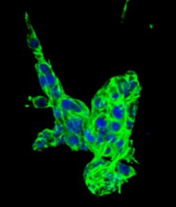
D2A1 Cells
Dr. Shira Michaeli/ Dr. Dalit Barkan Lab
Confocal image of D2A1, tumor cell line, outgrowth in the 3D BME system. Cells were treated with conditioned media for 7 days. F-actin filaments (green) and Nuclei (blue).